Skin Disorders
CLICK ON A TOPIC BELOW TO LEARN MORE
Abscesses
An abscess is a pocket pus caused by bacterial infection secondary to puncture wounds, scratches or bites. An abscess in the skin feels hot, looks red, swollen and is painful. Diagnosis is afforded via physical exam, fine needle aspirate and cytology. Treatment may include drainage, antiseptic flushing and antibiotics.
Acanthosis Nigricans
Acanthosis nigricans is a skin condition that appears as darkened skin in the underarm area of the front legs. As it progresses, the affected skin may thicken, fold, become greasy and produce a unpleasant odor. The condition occurs almost exclusively in Dachshunds. Treatment is palliative.
Acne
Acne is a skin disease consisting of comedomes (blackheads), papules (small reddened nodules) and pustules. In dogs, the disorder usually resolves with sexual maturity. In cats, the disorder is secondary to uncleaned chins. Diagnosis is via physical exam. Treatment consists of antiseptics, antibiotics and follicular cleansing (eg. benzoyl peroxide).
Acral Lick Dermatitis
Acral lick dermatitis is a skin condition secondary to repeated licking. Causes include boredom and stress. Signs include a small area of hair loss, progressing to a thickened, raised plaque which may become raw, inflamed and ulcerated. Dobermans, Great Danes, Labrador Retrievers, Irish Setters and German Shepherds are more likely to develop the condition. Diagnosis is afforded via history, physical exam, cytology and biopsy. Treatment may include behavior modification and anti-inflammatories.
Allergic Contact Dermatits
Allergic contact dermatitis is an itchy skin disorder that occurs in the thin-skinned areas of the body that contact the floor or ground when your pet lies down. The abdomen, chest, scrotum, ears and chin can also be affected. Secondary bacterial infections may occur. Signs may include redness, hair loss, pruritis (itchiness) and pustules. Diagnosis is afforded via physical exam, accurate history, cytology, biopsy and response to treatment. Treatment may include antiseptic cleansers, corticosteroids and antibiotics.
Allergies
Excessive scratching may indicate allergies. Allergic skin disease comes in a variety of forms such as inhalant dermatitis (atopy), contact dermatitis, flea bite allergy and food allergy. Some pets have occasional problems, others continually and still others may itch only at certain times or seasons of the year. Pets may be allergic to various things such as flea bites, pollens, molds, grasses, trees, house dust, wool, tobacco smoke, certain foods, and even other pets. Regardless of the allergen, the main signs are scratching and chewing of the skin. This action may result in extensive skin damage and secondary bacterial infection.
Atopy
Atopy is an intensely itchy skin disorder of both dogs and cats caused primarily by inhaled allergens (eg. house dust, pollen, molds). Allergens may also be absorbed through the skin. Atopy is an inherited disorder. Certain breeds of dogs are known to be genetically predisposed to the condition (eg. Golden Retrievers, Schnauzers, Lhasa Apsos, Labrador Retrievers, Bulldogs, Dalmatians, English Setters, Chinese Shar Peis, Irish Setters, Boston Terriers, West Highland White Terriers, Cairn Terriers and Wire-Haired Fox Terriers). The onset of atopy is usually between 1 and 3 years of age. Female dogs have a slightly higher rate of occurrence than males. Signs include itching, red skin, hair loss, face rubbing, foot chewing, inflamed ears, skin infection and hyperpigmentation. Diagnosis is afforded via accurate history, physical exam, blood testing and intradermal skin testing. Treatment may include skin cleansing, antibiotics, antihistamines, corticosteroids, omega 3 fatty acids, phytosphingosine and immunotherapy (allergy shots). Newer treatment includes oclacitinib (Apoquel) and lokivetmab (Cytopoint). Both are safe and very effective.
Bacterial Hypersensitivity
Bacterial hypersensitivity is an uncommon skin allergy elicited by certain bacteria (eg. Staphlococcus). Signs include pruritis, red sores, hair loss and scaling. Diagnosis is afforded via accurate history, physical exam, biopsy and immunological testing. Treatment may include topical therapy, corticosteroids, antibiotics and immune therapy (hyposensitization).
Calluses
Calluses are areas of thickened, hairless skin that develops in response to pressure over a boney protuberance. Common areas affected include the elbows and hocks of heavy pets. Calluses may be unsightly but are generally harmless. In rare cases the callus may become irritated, infected and/or fluid filled (hygroma) requiring medical and/or surgical treatment.
Cutaneous Cysts
Cutaneous cysts (eg. Dermoid and Epidermal Inclusion Cysts) are abnormal sac-like structures of the skin that develop due to displacement of skin forming cells (Dermoid)or obstruction of hair follicles (Epidermal). Most cysts appear as a small bump or nodule and contain a grayish, cheesy material. They may be multiple and become inflamed. Diagnosis is afforded by physical exam and aspiration. Treatment is via surgery.
Dermatophytosis (ringworm)
Dermatophytosis is a fungal disease of the skin commonly called ringworm. The fungus grows on the skin in dead skin cells and spreads out in a circular, reddened, crusty pattern. Fungi can be found in soil, on pets, on wildlife and on people. Pets may contract dermatophytosis from any of these sources and then pass it to others. Some pets may be nonsymptomatic carriers of the fungus. Diagnosis is afforded via physical exam, skin scraping, cytology and Dermatophyte Test Medium (DTM). Treatment may include hair clipping, topical antiseptics and antifungals (topical and/or oral). Avoid exposure of the infected pet to others. Wash your hands after handling the pet.
Ear Flap Alopecia (pinnal alopecia)
Pinnal aloecia is hair loss on the ear flaps. Causes include heredity (Dachshunds), metabolic disorders or inflammatory disease. Diagnosis is afforded via physical exam, laboratory tests and biopsy. Treatment is directed at any underlying disorders.
Ear Flap Dermatitis
Ear flaps are commonly irritated by biting insects such as black flies, mosquitoes and gnats. The resulting dermatitis may produce redness, hair loss, oozing serum, pruritus and secondary infections. Diagnosis is via physical exam and history. Treatment may include antiseptic cleansers, anti-inflammatories, antibiotics and insect repellents.
Endocrine Alopecia
Endocrine alopecia is a skin disorder secondary to hormonal imbalance that is characterized by nonpruritic, symmetrical hair loss (alopecia) involving the perineum, abdomen and inside of the thighs. Diagnosis is afforded via physical exam and history. Treatment may include hormonal replacement therapy.
Eosinophilic Granulomas
Eosinophilic granulomas are well demarcated, pruritic, raised sores of the lips, thighs and/or abdomen of cats. Causes include allergies, fleas, bacteria, fungi and viruses. Diagnosis is afforded via physical exam, skin scapings, cytology, culture, sensitivity, complete blood count and biopsy. Treatment may include corticosteroids, antihistamines, hyposensitization therapy, parasiticidal therapy, antibiotics and progesterone.
Flea Allergy Dermatitis
Flea allergy dermatitis (FAD) is a hypersensitivity reaction to components of flea saliva. Signs include pruritis, hair loss, scaling and hyperpigmentation along the tail base, rump and back of the hind legs. Diagnosis is afforded via accurate history, physical exam, biopsy, in vitro testing and intradermal testing. Intradermal testing is by far the gold standard for diagnosis. Treatment may include corticosteroids, antihistamines, omega 3 fatty acids, flea control (lufenuron), flea treatment (eg. imidacloprid, fipronil, selamectin, nitenpyram) and hyposensitization.
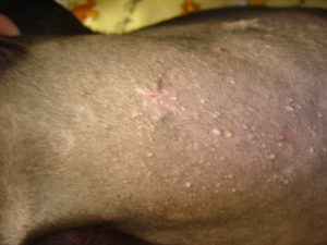
Folliculitis
Folliculitis is an inflammation of the hair follicle. The inflammation may be infectious or non-infectious. Infectious folliculitis can be caused by bacteria (eg. Staphylococcus), fungi or parasites (eg. Demodex). Non-infectious folliculitis is usually due to immune mediated reactions. Signs include small bumps at the base of hairs, redness and pruritis. Diagnosis is afforded via physical exam and histopathology. Treatment is directed toward the etiology. This may include antibiotics, anti-fungals and/or anti-parasitics. Non-infectious folliculitis may be treated with corticosteroids.
Food Allergy
Food allergy is a hypersensitivity reaction to food. Many affected pets have ingested the offending food substrate for over 2 years. Signs include red itchy skin, various skin lesions and in some cases, vomiting, diarrhea, respiratory distress, malaise or even seizures. Diagnosis of food hypersensitivity is difficult and time consuming. It requires feeding a hypoallergenic diet for at least 6-8 weeks and once symptom free, gradually adding back foods substances. Those re-initiating symptoms are eliminated from the pet’s diet. Treatment is avoidance of offending food substrates.
Adverse food reactions (Food allergies) in dogs and cats Characteristics:
- No sex predilection
- No correlation with a change of diet
- Non seasonal (ie. not related to one particular season)
- Age of onset is variable (4 months to 14 years)
- Causes pruritis (itchiness) of various parts of the body
- May present as a gastrointestinal disturbance (eg. Intermittent vomiting or diarrhea)
Diagnosis:
- Only accurate diagnosis is based on elimination diet leading to resolution of clinical signs (eg. pruritis) followed by reaction to reintroduced diet components.
- Elimination diets should be fed for a minimum of 8 weeks (no cheating!)
- Elimination diets are comprised of proteins and carbohydrates novel to the pet (ie. the pet has NEVER eaten this protein) with no additives (ie. artificial flavors, dyes, texturizers, preservatives, neoantigens, leachates, metals, etc.).
- Alternatives to elimination diets are hydrolysate diets (ie. commercially hydrolysed proteins such as found in HA by Purina or z/d by Hill’s)
NOTE: Prolonged feeding of a single protein source may increase the likelihood of developing an adversed reaction to that particular protein.
Treatment:
- Once the offending proteins and carbohydrates have been identified, that pet can NEVER eat that food source again! Be aware that the offending products may be present in treats, medications, vitamins, etc.
- Occasionally a novel protein given as a canned protein (and therefore cooked with carbohydrates in the canning process) may become an allergen when previously deemed OK. This is called a Maillard reactant product.
Hygroma
Hygroma of the elbow is a fluid filled sac that forms over the boney projections of the elbow and/or hock. It is caused by repeated trauma, pressure to the area in contact with hard surfaces. The condition is found most frequently in large breeds of dogs such as the Saint Bernard, Great Dane, Irish Wolfhound and Mastiff. It develops when the dog is relatively young, yet older animals may be affected. The swelling may become painful and ulcerated. Diagnosis is via physical exam. Treatment may include emollients, decubital blankets (ie. irregular surface), anti-inflammatories, antibiotics and surgery.
Interdigital Pyoderma
Interdigital pyoderma is an infection of the feet, between the toes. The condition is caused by bacteria secondary to allergies, trauma or irritants. Signs include pustules, draining sores, licking, redness, pruritis and hair loss. Diagnosis is afforded via physical exam, cytology, biopsy, culture and sensitivity. Treatment may include antibiotics, anti-inflammatories, antihistamines, omega 3 fatty acids, antiseptic cleansers and surgery.
Keratinization Defects
All of the following conditions are defects of keratinization manifested by localized or generalized excess scale formation.
Canine Acne
Fairly common disorder of follicular keratinization which results in comedones (blackheads), bacterial folliculitis and furunculosis.
Ear Margin Dermatosis
Rare bilaterally symmetric keratinization defect of the ears of Dachshunds.
Epidermal Dysplasia
Severe scaling disorder of West Highland White Terriers.
Ichthyosis
Extremely rare congenital (present at birth) keratinization defect of Terriers.
Idiopathic Nasodigital Hyperkeratosis
Excess keratinization disorder of the nose and footpads. Seen mostly in Cocker Spaniels and English Spaniels.
Lichenoid-Psoriasiform Dermatosis
Extremely rare keratinization defect of English Springer Spaniels.
Primary Idiopathic Seborrhea
The most common keratinization disorder of dogs ranging from dry to greasy scaling. Predisposed breeds include Cocker Spaniels, English Springer Spaniels, West Highland White Terriers, Basset Hounds, Irish Setters, German Shepherds, Dachshunds, Doberman Pinschers, Chinese Shar Peis and Labrador Retrievers.
Schnauzer Comedo Syndrome
Follicular keratinization defect of Miniature Schnauzers producing black heads (comedones) along the midline of the back.
Sebaceous Adenitis
An inflammatory disease of the sebaceous (oil) glands of Standard Poodles, Akitas, Samoyeds and Vizslas whereby keratinization defects obstruct the sebaceous ducts.
Vitamin A-Reponsive Dermatosis
Rare scaling disorder which responds to high doses of topical Vitamin A primarily seen in Cocker Spaniels. (Also seen in Miniature Schnauzers, Labrador Retrievers and Chinese Shar Peis.)
Zinc Responsive Dermatosis
Rare scaling disorder of Alaskan Malamutes and Siberian Huskies which responds to zinc supplementation.
*Diagnosis of keratinization disorders is afforded via history, breed, diagnostic elimination of secondary causes of scaling and skin biopsy. Treatment is directed at controlling scale formation, supplementing appropriate nutrients and corticosteroids if appropriate.
Nasodigital Hyperkeratosis
Nasodigital hyperkeratosis is a disorder characterized by excessive growth of the epithelium of the nose and footpads. The cause is unknown. The nose and pads thicken, harden, dry and crack. Diagnosis is afforded via physical exam and biopsy. Treatment may include emollients, topical vitamin A, corticosteroids and debridement.
Pemphigus Complex
Pemphigus complex is a group of severe immune mediated skin diseases. Signs include skin lesions such as redness, blisters, scaling and crust around the face, mouth, nose, anus and/or genitalia. Diagnosis is afforded via physical exam, complete blood count, immune tests, cytology and biopsy. Treatment may include corticosteroids, immune suppressive therapy and supportive therapy.
Perianal Fistula
Perianal fistula is an abnormal opening in the skin near or around the anus. The fistulas may reach deep into the muscle sheaths of the pet’s hind end. Infection is common and the condition is painful. Signs include draining tracts, odor, redness, pain and a reluctance to raise the tail. Diagnosis is afforded via physical exam, radiography with dye, culture and sensitivity. Treatment may include antibiotics, cyclosporine, anti-inflammatories, and cryosurgery, laser surgery or conventional surgery. Tail amputation is usually recommended in breeds with low set tails such as German Shepherds.
Psychogenic Dermatitis
Psychogenic dermatitis is a disorder of cats characterized by intense licking of a local skin area. The cat’s rough tongue erodes the skin exposing sensitive nerve endings, stimulating the cat to lick even more, producing more damage and a vicious cycle of licking, damage, licking, etc. Causes include infection, allergies, trauma or simply due to an anxiety precipitated by a change in environment, surroundings, boarding, loss of a companion, threats by a neighboring cat or insensitivity of a family member. Signs include hair loss and extensive skin damage. Psychogenic dermatitis is more common in Siamese and Abyssinian cats. Diagnosis is afforded via accurate history, physical exam, complete blood count, blood chemistries, culture, sensitivity, skin scraping, intradermal skin testing, laboratory tests, biopsy and video taping of the cat’s activity and interaction over an entire day or more. Treatment may include anti-anxiety medication, corticosteroids, treatment of secondary skin lesions and elimination of underlying causes.
Pyoderma
Pyodermas are skin infections caused by pus-producing bacteria. Pyodermas may involve only the outermost skin layers (superficial pyoderma), deeper skin layers (deep pyoderma) or both. Signs include redness, pustules, pruritis, hair loss and odor. Diagnosis is afforded via physical exam, culture, sensitivity, skin scraping, cytology and biopsy. Treatment may include anti-inflammatories, topical antiseptics, shaving, frequent shampooing and antibiotics. These infections require rigorous treatment for a prolonged period. Pyodermas can be especially difficult to treat. Occasionally the infections are highly resistant to antibiotics, making effective medication difficult. Repeated cultures of the skin may be necessary.
Schnauzer Comedo Syndrome
Schnauzer comedo syndrome is characterized by development of comedones (blackheads) in the skin of Miniature Schnauzers. Cause is a keratinization disorder of the hair follicles. The blackheads are most numerous on the back, from the neck to the pelvis. Comedones appear as hard, crusted projections on the skin surface. Diagnosis is afforded via physical exam and possibly biopsy. Treatment may include benzoyl peroxide shampoos, gentle agitation with a mildly abrasive sponge, antibiotics and synthetic retinoids (eg. isotretinoin).
Seborrhea
Sebum is a normal product of skin sebaceous glands. In seborrhea, excessive sebum is produced and may appear as dry, light-colored flakes or as greasy, waxy scales on the skin and hair. Sebum is a fatty material that becomes rancid and causes a strong coat odor. Seborrhea may occur by itself (primary seborrhea) or result from a underlying disease (secondary seborrhea). The cause of primary seborrhea is unknown (primary idiopathic seborrhea). Secondary seborrhea often clears up when the underlying disease is cured, while primarily seborrhea is a chronic disease that may be controlled but not cured. Diagnosis is afforded via accurate history, physical exam, skin scraping, complete blood count, blood chemistries, laboratory tests, culture, sensitivities, cytology and biopsy.
Skin and Coat Care
By following basic guidelines, you can easily care for your pet’s skin and coat:
- The skin and coat reflect your pet’s general health. Healthy pets have fewer skin and coat problems. Visit your veterinarian regularly with pet health problems as well as for annual pet health exams.
- Avoid over the counter, antidotal or exotic health remedies. Consult your veterinarian.
- Parasites (fleas, intestinal parasites, etc.) affect the skin and coat. Follow your veterinarian’s suggestions for parasite control.
- Proper nutrition is essential for healthy skin and coat. Discuss your pet’s diet with your veterinarian.
- Routine grooming prevents skin and coat problems, while enabling you to detect problems early in their progression.
- When bathing your pet, use a mild pet shampoo, rinse well, towel off excessive water and blow dry with a hair dryer while brushing.
- Long-haired pets should be combed daily without fail. Short-haired pets should be brushed at least once a week. Special attention should be given to areas where mats are likely to form (ie. behind the ears, under the legs, on the stomach, back legs and under the tail).
Solar Dermatitis
Solar dermatitis is sunburn of the ears in pets. Sun exposure results in reddening of the skin and thinning of the hair on the tips of white ears. With continued exposure, skin peels, crust forms and the ears begin to itch. Damage may continue from continued sun exposure, the pet’s scratching and secondary skin infections. Skin cancer (squamous-cell carcinoma) may develop in the damaged areas. Diagnosis is afforded via history, physical exam and skin biopsy. Treatment may include sun block, avoidance of direct sun exposure, treatment of secondary lesions and surgery.
Stud Tail in Cats
Cats have a multitude of skin glands at the tail base. Especially in confined cats, glandular secretions accumulate and cause fur matting with a scaly, crusty accumulation. Since the condition is most common in uncastrated males, the condition has been dubbed “stud tail.” Nonetheless, neutered male and female cats may be affected. Castration does not correct this problem.


