Reproductive Disorders
CLICK ON A TOPIC BELOW TO LEARN MORE
Acute Metritis
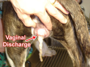
Acute metritis is a rapidly developing infection of the uterus. It can result from abortion, retained placenta or mild uterine infection (eg. secondary to owner assisted births). Signs include vaginal discharge (with blood and pus), fever, depression, loss of appetite and refusal to care for young. Offspring often cry excessively and seem restless even after nursing. Acute metritis requires immediate medical attention. Most affected pets should be spayed.
Agalactia
Agalactia is the lack of milk production in a nursing mother. There is no medication to increase milk production but the hormone, oxytocin, can stimulate release of milk from the mammary glands.
Artificial Insemination
Artificial insemination is feasible if two dogs are incompatible for some reason or when either dog is sexually inexperienced. The procedure is safe and conception rates are good when performed under proper conditions by a qualified veterinarian.
Artificial insemination involves 3 basic steps:
- Detecting the time of ovulation by vaginal cytology, rising progesterone level and/or behavioral response to the presence of a male.
- Collection of semen from the male dog.
- Insemination of the female dog.
Balanoposthitis
Balanoposthitis is an inflammation of the penis and prepuce. Causes include bacteria, herpesvirus, fungi, trauma, foreign bodies and neoplasia. Signs include discharge (pus or blood), discomfort, excessive licking, urinary incontinence, urination in abnormal direction, unwillingness to copulate or no signs. Diagnosis is afforded via complete history, physical exam, imaging studies, cytology, culture and sensitivity. Treatment may include antibiotics, surgery, antifungals and povidine-iodine douches.
Care of the Newborn and Mom
Pregnancy and nursing offspring constitute a severe strain on the mother’s health. Though most mothers handle the task well, the owner should take certain precautions to protect the health of both mother and offspring.
Important Points in Postnatal Care
Physical Examination: Within 48 hours after birth, the mother and offspring should be examined by a veterinarian.
Diet: The mother should receive a highly digestible, balanced diet. Calories should be increased by 20% per nursing offspring. This can be accomplished by offering growth diets (eg. puppy or kitten foods). Additional calcium can be added via supplementation.
Fluids: Provide clean, fresh water at all times.
Activity: Ordinarily the mother will spend most of the first few weeks with her offspring. Allow her any exercise as she desires.
Bowel Movements: Your pet may have to relieve herself more frequently. Stools may be soft for awhile but if diarrhea or straining develop, contact your veterinarian.
Mammary Gland Care: Check the nipples daily and clean with warm water if debris is present. Inform your veterinarian to any discoloration of the skin, swelling, tenderness or sores. Trim the offspring’s nails if they are scratching the mammary glands.
Vaginal Discharge: A reddish vaginal discharge, with clotted blood, is normal for the first few days postpartum and may occur intermittently for several weeks.
Behavior: Call the doctor if the nursing mother appears uncomfortable or refuses to nurse the puppies.
General Effects: Normally the mother experiences heavy coat shedding and slight weight loss during the nursing period. Brush her regularly and call the doctor if the mother appears overly thin.
Estrus (heat period): Pregnancy usually has no effect on the next heat period. This period should occur within 6 months after birth of the puppies or at the next season for cats.
Spaying (ovariohysterectomy): If you desire surgical sterilization for your pet, an appointment should be scheduled after the puppies or kittens are weaned and milk production has ceased, but before the next heat period.
Cesarean Section
A cesarean section is a procedure to surgically remove puppies or kittens from the uterus when the mother cannot deliver the offspring or natural (unassisted) delivery is potentially harmful to the mother or babies. Reasons for cesarean section are numerous and include mother’s illness, mechanical obstructions in the birth canal (healed fractures, tumors or abdominal masses) or unusually large offspring. Surgery may be planned well in advance because of known problems or performed only after difficulties develop at the time of birth. After cesarean section, the mother can usually assume her normal maternal duties. The surgical incision and sutures rarely interfere with nursing but if problems arise, consult your veterinarian.
Cryptorchidism
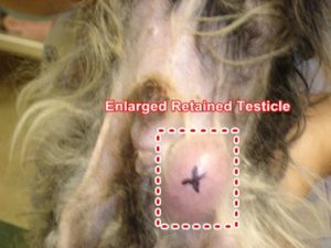
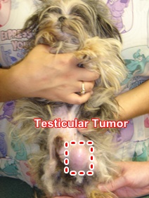
During development of the male fetus, testicles develop in the abdominal cavity. The testicles pass through an opening in the body wall called the inguinal canal and descend into the scrotum. In some individuals, one or both testicles fail to descend into the scrotum. Pets with both testicles undescended are usually sterile while those with only one undescended testicle are fertile. Undescended testicles are more common in small or toy breeds. Pets with undescended testicles should be castrated, since the condition is hereditary and the incidence of testicular tumors in undescended testicles is over 10 times greater than in descended testicles.


Endometritis
Endometritis is an inflammation/infection of the uterine lining. The disorder usually causes no signs of illness other than infertility. These pets usually cycle and mate normally but conception rates are very low. Some pets with endometritis develop a more serious infection called pyometra. They may become very ill and vomit, drink large amounts of liquids, urinate frequently and have a vaginal discharge. Untreated pyometra can cause death. Diagnosis is afforded via cytology, culture and sensitivity of the cervix. Treatment may include antibiotics and ovariohysterectomy.
Estrus
Estrus (“heat”) is the mating period of female animals. When estrus occurs, animals are said to be “in heat” or “in season”.
Cats normally have their first estrous cycle between 5 and 10 months of age (average 6 months). The female cat has 2-4 estrous cycles every year, each lasting 10-20 days. If she is bred, estrus seldom lasts more than 5 days. If not bred, estrus may last for 7-10 days and recur in 14-21 days. An unmated female may cycle every 3-4 weeks if greater than 14 hours of light is provided each day. Cats cycle again 1-6 weeks after giving birth, so a female may be nursing one litter while pregnant with another.
Dogs generally have their first estrous cycle at 6-12 months of age. Some large breeds may not have their first estrus until 12-24 months of age. The complete cycle takes approximately 6-12 months (average 8 months), resulting in 1-2 estrous cycles each year. Individual variation occurs, but a given female’s cycle tends to be regular.
The estrous cycle can be divided into 4 stages:
- Proestrus: This stage begins with vaginal swelling and bleeding. It normally lasts from 7-10 days. Male dogs become very interested in the female; however, she remains antisexual.
- Estrus: In this stage, the female will accept the male and conception can occur. Vaginal discharge is more yellowish than bloody. Ordinarily, the stage lasts 7-10 days. The female will stand still and hold her tail to the side when touched on her back or a male dog tries to mount.
- Metestrus: In this stage, the corpus luteum (part of ovary that helps maintain pregnancy) forms and is functional. False pregnancies frequently occur during this stage.
- Diestrus: In this stage, sex organs are quiet.
Galactostasis
Galactostasis is a disorder that occurs when mother’s milk is so abundant the offspring cannot suckle enough to empty the mammary glands. As a result, the milk tends to harden in any mammary glands not suckled empty by offspring. The mammary glands become reddened and painful. Mastitis (inflammation of the mammary glands) is a common sequella.
Gestations
The term gestation means the period when the young are developing within the uterus. Normal gestation in cats is 63-65 days. Siamese cats may carry their kittens for 67 days.
Normal gestation in dogs is 63 days. Due to variation in actual conception relative to mating, puppies may be delivered between 58 and 70 days after a single mating.
Hypocalcemia in Lactation
Hypocalcemia (milk fever) occurs most frequently in smaller breeds. Females with heavy milk production and large nursing litters are most likely to be affected. The cause appears due to an imbalance between calcium uptake from the digestive tract and calcium outflow in milk, bone, urine and feces. Calcium replacement is vital since lack of proper treatment can be fatal. Milk fever may recur in subsequent pregnancies, therefore future prevention is imperative.
Infertility
Infertility is the inability to reproduce. Causes are complicated and may include general health, age (too young or too old), disease of the reproductive organs, endocrine (hormone) disorders, stage of the female sexual cycle (heat), emotional state (shyness, fear, excitement), and the environment at breeding (unfamiliar surroundings, distractions, noises and strangers). Your veterinarian may examine vaginal smears and sperm cytology to determine optimum breeding time and capability.
Mastitis
Mastitis is an inflammation/infection of the mammary glands related to heavy milk production and incomplete emptying of the glands. Signs include fever, restlessness, reduced appetite, swollen, tender and hot mammary glands, dark-red or purple soft spots and blood-tinged or off-color milk. The mother will often neglect the offspring, who if not cared for will cry excessively, weaken and die.
Mating in Cats
Cats experience their first estrus cycle at approximately 7 months of age. They cycle every 2-4 week in the spring or with 14 hours of light per day. Each estrus lasts approximately one week. During the estrus cycle, cats may act “strangely” (eg. vocalize, raise their tails with pressure on the rump). Prior to mating, both cats (male and female) should be examined by your veterinarian and tested negative for feline leukemia, feline immunodeficiency virus, external parasites and contagious disease. Place the breeding pair together in a large, clean area for up to a few hours until copulation has been witnessed several times. Be sure the cats are compatable.
Mating in Dogs
Female dogs generally have 2 reproductive cycles each year. Small breeds first cycle at approximately 5-6 months of age, while some giant breeds may not cycle for the first time until 2 years of age. The average age at puberty is approximately 6-10 months. Female dogs should not be bred during the first heat period due to immaturity. Instead, wait until at least the second heat to breed the female. At the beginning of the female cycle there is a bloody discharge for about 7-10 days. After about 4-8 days of bloody vaginal discharge, the female will accept the male and stand for breeding. This receptive period may last a few days or as long as 2 weeks. Commonly used breeding dates are the 9th, 11th and 13th days after the first vaginal discharge. Repeated breedings every 48 hours, for as long as the female accepts the male, produce the best conception rate. Assistance is rarely needed for a successful mating, especially if the dogs have had previous experience. Occasionally some assistance must be given such as with the male mounting and entering the female or restraining the female so as not to harm the male. A muzzle (gauze, nylon stocking, etc) tied around the female’s muzzle may be helpful. With normal mating, the dogs become “tied” together for up to 1/2 hour. Occasionally, the male turns 180 degrees around and the dogs appear “end to end”. This is normal but if the dogs become over active during this stage, gentle restraint is advisable. Do not attempt to pull the dogs apart, since this may cause injury. Trouble with breeding may indicate the timing is not correct. Review dates and consult with your veterinarian.
Pregnancy represents a considerable strain on the mother and therefore females should not be bred every “season”. Appropriate breeding programs breed every other heat. With successful mating, puppies should be born 63-66 days from the first breeding.
If you are considering mating your dog, discuss the matter ahead of time with your veterinarian. A thorough examination of both the male and female, including Brucellosis testing, is recommended before breeding to help ensure that the pets are in good physical condition and free of contagious disease.
Normal Birth in Cats
Prepare for delivery of kittens before the female gives birth. Provide a queening box just large enough for the mom to stretch out in, with sides 6-8 inches high. This will keep the kittens from crawling out of the nest. Place the box in a comfortable, secluded area of the home, away from the family traffic. Keep the bedding clean and dry (eg. shredded paper, cloth, blankets, etc.)
Within 24 hours before the onset of labor, the teats will normally fill with milk. Labor in the female cat can be divided into 3 stages; the last two repeating with the birth of each kitten.
Stage 1: The female appears restless, often seeks seclusion and may refuse food. This stage may last approximately 6-24 hours. Allow access within the house for the cat to urinate, defecate and exercise.
Stage 2: In the second stage, contractions and expulsion of the kittens begin. Usually a small sac of fluid protrudes first from the vulva, followed by the kitten and its attached placenta. The kitten normally presents nose first, yet some kittens are born hindquarters first. Both presentations are considered normal in the cat. After delivery, the mother tears off the sac, cleans up the kitten and severs the umbilical cord. Make sure the sac is removed from the kitten immediately if it is unbroken and still on the offspring’s face post delivery.
Stage 3: In the third stage of labor, mild contractions and delivery of the afterbirth take place. The mother rests between births. This stage may range from a few seconds to an hour.
If the mother is disinterested in the kittens or inexperience precludes her from proper care, remove all membranes covering the kitten; clean the face and remove mucus from the mouth and nose; rub and dry the kitten with a clean towel; and stimulate respiration and circulation. After a few minutes the kitten should begin to squirm and cry out loud. If the placenta is still attached, tie off the umbilical cord about an inch from the kitten’s body with fine thread and then cut on the side of the knot away from the kitten. Apply a drop of iodine or triple antibiotic ointment to the cord end after it is cut. If a kitten seems to be lodged in the birth canal and the mother cannot expel it, rapid assistance may be necessary. Call your veterinarian immediately. At home, assistance can be afforded by grasping the kitten with a clean towel and exert steady, firm traction without jerking or pulling suddenly. Traction may have to be applied for several minutes. If the kitten cannot be removed, transport the mother and kitten to your veterinarian immediately.
During queening and nursing, your pet may be very nervous, protective and slightly aggressive. Her attitude should normalize with time, so be cautious.
Notify the doctor if any of the following occur:
- A lodged kitten cannot be readily removed from the birth canal.
- Strong, persistent labor lasts for greater than 30 minutes without delivery of a kitten.
- Weak, intermittent labor lasts for 6 hours without delivery of any kittens.
- More than 4 hours elapses since the last birth and it is probable that more kittens are still inside.
- A watery discharge presents with no labor or kittens within 3-4 hours.
- Pregnancy lasts more than 68 days.
Normal Birth in Dogs
Prepare for delivery of puppies before the female gives birth. Provide a whelping box just large enough for the mom to stretch out in, with sides 6-8 inches high. This will keep the pups from crawling out of the nest. Place the box in a comfortable, secluded area of the home, away from the family traffic. Keep the bedding clean and dry (eg. shredded paper, cloth, blankets, etc.)
Delivery time can be more precisely predicted by monitoring the dog’s rectal temperature and teats. Check the rectal temperature of the mother twice daily from the 58th day of pregnancy until labor begins. Normal rectal temperature varies between 99.5o to 102.5o F. Within 24 hours before the onset of labor, the rectal temperature drops approximately 1-2 degrees. The teats will normally fill with milk approximately 24 hours prior to delivery. Labor in the female dog can be divided into 3 stages; the last two repeating with the birth of each puppy.
Stage 1: The female appears restless, often seeks seclusion and may refuse food. This stage may last approximately 6-24 hours. Exercise the dog to allow for urination and defecation.
Stage 2: In the second stage, contractions and expulsion of the puppies begin. Usually a small greenish sac of fluid protrudes first from the vulva, followed by the puppy and its attached placenta. The puppy normally presents nose first, yet some puppies are born hindquarters first. Both presentations are considered normal in the dog. After delivery, the mother tears off the sac, cleans up the pup and severs the umbilical cord. Make sure the sac is removed from the puppy immediately if it is unbroken and still on the offspring’s face post delivery.
Stage 3: In the third stage of labor, mild contractions and delivery of the afterbirth take place. The mother rests between births. This stage may range from a few seconds to an hour.
If the mother is disinterested in the puppies or inexperience precludes her from proper care, remove all membranes covering the puppy; clean the face and remove mucus from the mouth and nose; rub and dry the puppy with a clean towel; and stimulate respiration and circulation. After a few minutes the puppy should begin to squirm and cry out loud. If the placenta is still attached, tie off the umbilical cord about an inch from the puppy’s body with fine thread and then cut on the side of the knot away from the puppy. Apply a drop of iodine or triple antibiotic ointment to the cord end after it is cut. If a puppy seems to be lodged in the birth canal and the mother cannot expel it, rapid assistance may be necessary. Call your veterinarian immediately. At home, assistance can be afforded by grasping the puppy with a clean towel and exert steady, firm traction without jerking or pulling suddenly. Traction may have to be applied for several minutes. If the puppy cannot be removed, transport the mother and pup to your veterinarian immediately.
During whelping and nursing, your pet may be very nervous, protective and slightly aggressive. Her attitude should normalize with time, so be cautious.
Notify the doctor if any of the following occur:
- A lodged puppy cannot be readily removed from the birth canal.
- Strong, persistent labor lasts for greater than 30 minutes without delivery of a pup.
- Weak, intermittent labor lasts for 6 hours without delivery of any puppies.
- More than 4 hours elapses since the last birth and it is probable that more puppies are still inside.
- A greenish-black discharge presents with no labor or puppies within 3-4 hours. The greenish-black color is normal, but should rapidly be followed b
Orchitis
Orchitis is inflammation and/or infection of the testicles. Causes include testicular torsion, infection (eg. from the urinary bladder, prostate gland or bloodstream), Brucella canis, sperm granuloma, varicocele (dilation and thrombosis of the spermatic vein), neoplasia, immune-mediated disease, toxins and trauma. Signs include pain, swelling, licking of the scrotum, anorexia, listlessness, fever, reluctance to walk, stiff gait and possibly abdominal pain. Diagnosis is afforded via physical exam, ultrasound, semen evaluation, cytology, biopsy, complete blood count, culture and sensitivity. Treatment may include antibiotics, antifungals, anti-inflammatories and castration.
Phimosis/Paraphimosis
Phimosis is the inability to protrude the penis through the opening of the sheath. Causes include a congenital (present at birth) abnormally small opening in the sheath or scarring of the opening as a result of injury or disease. Phimosis can result in abnormal urination and distention of the sheath.
Paraphimosis is the inability to withdraw the penis back into the sheath. This disorder often occurs in young dogs after mating, injury or masturbation. Causes also include an abnormally small sheath opening or hair on the sheath that folds inward and traps the penis outside.
Diagnosis of these disorders is afforded via physical exam. Treatment may include anti-inflammatories, corticosteroids, topical therapy and surgery.
Prostatic Hyperplasia
The prostate gland surrounds the tubular outflow tract (urethra) of the urinary bladder in male dogs. It produces most of the fluid portion of semen. Prostatic hyperplasia is the abnormal enlargement of the prostate gland. It is associated with a hormone imbalance and is common in intact (non-castrated) dogs over 5 years of age. The enlarged prostate compresses the colon (located directly above the prostate) which can cause difficult, painful bowel movements and eventually constipation. Other signs include blood in the urine, blood dripping from the prepuce, difficult urination, diarrhea and chronic urinary tract infections. Diagnosis is afforded via physical exam (include digital rectal exam), imaging studies, urinalysis, prostatic fluid analysis and biopsy. Treatment may include anti-inflammatories, antibiotics, estrogen therapy, androgen inhibitors (eg. delmadinone acetate) and castration.
Prostatitis
The prostate gland surrounds the tubular outflow tract (urethra) of the urinary bladder in male dogs. It produces most of the fluid portion of semen. Prostatitis is a bacterial infection/inflammation of the prostate. The disorder is via bacterial contamination of the penis or blood stream which spreads to the prostate. Most male dogs are susceptible. Signs may include pain, blood in the urine, blood dripping from the prepuce, straining to urinate, difficulty defecating, depression, anorexia, vomiting, diarrhea and chronic urinary tract infections. Diagnosis is afforded via physical exam, digital rectal exam, imaging studies, complete blood count, urinalysis, prostatic fluid analysis and cytology, biopsy, culture and sensitivity. Treatment may include antibiotics, anti-inflammatories, support therapy and castration.
Pseudocyesis (false pregnancy)
False pregnancy is caused by a hormonal imbalance produced by the ovaries of a non-pregnant female 6-8 weeks after estrus. Signs may include restlessness, distended abdomen, lactating teats, nesting, guarding enclosed areas and “mothering” of toys, shoes or other articles. Diagnosis is afforded via accurate history, physical exam and determination that the female is not pregnant. Treatment is usually not necessary.
Pyometra
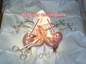
Pyometra is a bacterial infection with pus accumulation within the uterus. Pyometra results from hormonal influences that thicken the uterine lining and decrease the normal resistance to infection. As a result, bacteria enter the uterus through the open cervix of estrus resulting in infection. Closure of the cervix after infection can result in large accumulations of pus. Signs of pyometra may include loss of appetite, excessive thirst, depression, vomiting and fever, with or without vaginal discharge. The disease may develop slowly over several weeks. It is more common in older females but can occur in younger pets (especially if exposed to estrogenic therapy). Diagnosis is afforded via physical exam, complete blood count, blood chemistries, radiography, ultrasonography, cytology, culture and sensitivity. Treatment may include antibiotics, prostaglandin therapy (with “open”, draining pyometras), support therapy and ovariohysterectomy.

Termination of Pregnancy
Early, unwanted pregnancy is best ended by ovariohysterectomy (spaying). This irreversible operation removes both ovaries and the uterus. The pet is then permanently sterile and will no longer experience the reproductive “heat” cycle. If permanent sterility is not desired, hormone therapy can be administered to block uterine implantation of fertilized eggs. The eggs are then expelled with uterine secretions. In a small percentage of cases, hormonal termination of pregnancy may cause some undesirable side effects. Ask your veterinarian to discuss the risks and benefits of each type of treatment. Hormonal treatments are only successful if initiated within a few days after mating. Such treatment may extend the heat period. Males continue to be attracted and mating may occur although pregnancy should not take place.
Vaginal Hyperplasia
Vaginal hyperplasia is an exaggerated enlargement of the vagina. Vaginal hyperplasia occurs most commonly in young females during their first heat period and recurs with subsequent cycles. With each “heat” cycle the vaginal lining becomes congested and swollen as a result of high estrogen levels produced by the ovaries during this time. In some females, the swelling becomes excessive and vaginal tissue protrudes from the vaginal opening as a pinkish, fleshy mass. Most dogs recover spontaneously, yet others require treatment to prevent injury to the exposed vaginal tissue. Treatment may include surgical removal of the ovaries and uterus (spaying).
Vaginal Prolapse
ginal prolapse is a disorder whereby the vagina turns inside out and bulges through the vulva. Causes include hormonal stimulation of the vulva, straining during delivery or inappropriate human attempts to assist with delivery. The prolapse appears as a pink, fleshy mass that later may become dark red, bloody, infected and ulcerated. The pet may strain and frequently attempt to pass urine. Diagnosis is afforded via physical exam. Treatment includes surgery and supportive care. Your veterinarian can advise on future breeding.
Vaginitis
Vaginitis is inflammation of the vagina. Causes include developmental abnormalities (eg. vaginal stenosis, persistent hymen, vulvar hypoplasia), neoplasia, infection, foreign objects and mating injuries. Females may exhibit mild vaginitis before their first heat period (juvenile vaginitis). Juvenile vaginitis generally resolves after the first heat period. Signs of vaginitis vary with the cause and severity and may include vaginal discharge, excessive licking, frequent urination, attraction of males and redness. Diagnosis is afforded via physical exam, digital exam, culture, sensitivity, imaging studies and urinalysis. Treatment may include antibiotics, antiseptic douches and surgery.


