Alimentary Tract Disorders (GI tract, etc.)
CLICK ON A TOPIC BELOW TO LEARN MORE
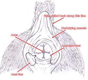
Anal Sac Disease
The anal sacs are located on each side of the anus, just under the skin with openings at 4 and 8 o’clock. They produce a foul smelling secretion which normally empties with each bowel movement. Diseases of the anal sacs can be categorized as either: impaction (thick fluid that can not be secreted) or infection (pus or chunky secretions which may even break through the skin). Signs of the disease include scooting (dragging the anus across the floor), excessive licking the anal area, and/or bloody discharge from the anal area. Physical exam (with anal sac expression) is used to diagnose the condition. Treatment includes expression, antibiotics (possible culture and sensitivity of the secretions), warm compresses and even surgical removal if the condition continues despite medical intervention.
Cleft Palate
Cleft palate is an inherited birth defect whereby the roof of the mouth has an abnormal opening to the nasal cavity. This allows food and liquid to enter the nose when feeding. The cleft may involve the hard palate, soft palate or both, and vary in size from a narrow slit to a defect encompassing nearly the entire palate. Larger defects can result in material entering the respiratory tract/lungs thereby producing aspiration pneumonia. Accurate history and a thorough oral exam help diagnose the condition. Diagnostic imaging can characterize concurrent aspiration pneumonia. Treatment is surgical correction. Very young pets may have surgery delayed until growth affords the surgeon additional tissue for closure. Stomach tube feeding should be implemented if surgery is delayed. This condition is heritable therefore animals with this condition should not be bred.
Colitis
Colitis is an inflammation of the large bowel (colon). Causes include parasites such as whipworms, bacteria, foreign bodies and food sensitivities or allergies. Common signs may include diarrhea, frank blood, straining, abdominal pain and/or dehydration. Complete blood counts, blood chemistries, laboratory tests, fecal exams, endoscopy, accurate history and abdominal imaging are used to diagnose the condition. Treatment is directed at both the etiology of the disease as well as its symptoms.
Constipation
Constipation is usually a sign of large bowel disorder and is characterized by infrequent and/or difficult bowel movements. Stools may be hard and dry causing pain and straining. Constipation is not a disease in and of itself. Diagnosis is attained through history and physical exam. Treatment is directed toward the underlying condition causing the constipation. Further diagnostics are likewise directed toward the underlying condition. Symptomatic treatment includes diet, laxatives and enemas.
Dental Disease
Dental disease begins with the accumulation of plaque (a clear combination of food particles, saliva and bacteria) upon the tooth surface down in the gingival sulcus (a canal between the tooth and gum). As plaque continues to accumulate, irritation of the gingiva (gums) produces inflammation (gingivitis). Gingivitis leads to bleeding of the gums. This bleeding mixes blood components with plaque producing tartar (calculus). As the process continues, the tartar works its way down the tooth root as well as out onto the visible surface of the tooth. Tartar and its contaminants can loosen teeth and spread infection leading to local abscess, as well as heart, liver, and kidney disease. Contaminants in contact with the lining of the mouth, can lead to tumor formation. Diagnosis of dental disease involves complete oral examination and dental radiographs. Treatment includes cleaning, polishing, root planing, gingival treatment and possible endodontic procedures.
Diarrhea
Diarrhea is a passage of fluid stool. It is not a disease itself, but a common sign of small and/or large bowel disease. Characteristics of diarrhea (fluidity, mucous, blood, frequency, gas) help in the proper diagnosis of small vs large bowel disease. Treatment is directed toward the underlying disease. History, fecal exams, diagnostic imaging, complete blood counts, blood chemistries, laboratory tests and endoscopy are used in the diagnosis of diarrhea etiology. Persistent diarrhea can lead to dehydration. Veterinary care is essential.
Enteritis
Enteritis is inflammation of the small intestine. Causes of enteritis include bacteria, viruses, fungi, parasites, immune disorders, food sensitivities, foreign material, metabolic disorders, etc. Complete blood counts, blood chemistries, laboratory tests (such as serum folate concentration, serum cobalamin concentration, fecal alpha-1-proteinase inhibitor concentration and serum C-reactive protein concentration), fecal exams, abdominal imaging, endoscopy and accurate history are utilized to diagnose enteritis and its etiology. Treatment is directed toward the underlying etiology.
Eosinophilic Gastroenteritis
Gastroenteritis is an inflammation of the stomach and small intestine. Eosinophilic refers to the type of white blood cell involved in the inflammation. Their presence is due to an immune mediated reaction to bacteria, parasites or another type of allergen (substances the body reacts to). The inflammation can result in vomiting, diarrhea, weight loss and/or dehydration. Complete blood counts, blood chemistries, laboratory tests, fecal exams, endoscopy with biopsy are utilized to diagnose this condition. Treatment may be prolonged and include immunosuppressive drugs and a hypoallergenic diet.
Gastric Dilation/Volvulus (bloat, gastric torsion)
Gastric dilation/volvulus is a life-threatening disease whereby the stomach balloons (dilation) with gas and may twist (volvulus), resulting in a closure of both the inflow and outflow tracts of the stomach. The swelling (with or without twisting) can block return of blood from the abdomen to the heart resulting in tissue congestion and eventual shock. Widespread tissue damage leads to kidney, respiratory and cardiac failure then death. This condition can affect any breed, but most cases involve older, large, deep-chested dogs. Causes are related to age, diet, activity in approximation to meals or ingestion of large amounts of water. Sign include anorexia, bloat, attempts to vomit, shock and death. Diagnosis is afforded via history, breed and age considerations, physical exam (including percussion exam), imaging studies (eg. X-rays), complete blood counts, blood chemistries and laboratory tests. Treatment may include immediate decompression, correction of electrolyte and blood gases abnormalities, cardiac treatment, surgical replacement of proper anatomy, surgical pexy (anchoring) of the stomach to prevent future displacements and dietary adjustments.
Gastritis
Gastritis is an inflammation of the stomach. Causes include ingestion of spoiled food, garbage, bones, foreign bodies, drugs, toxins, as well as infectious agents. Signs include vomiting and anorexia. Often the offending agent may not be identified. Bright and alert adult pets with no exposure to toxins should be kept from food and water for 4 to 6 hours. This allows the stomach to “rest”. Young pets, old pets, pets exposed to toxins or adult pets which continue to vomit should be examined by their veterinarian promptly. Complete blood counts, blood chemistries, laboratory tests, fecal exams, endoscopy, accurate history and abdominal imaging are used to diagnose the condition. Treatment is directed at both the etiology of the disease as well as its symptoms.
Gastroenteritis
Gastroenteritis is an inflammation of the stomach and small intestine. Causes include bacterial, viral or fungal infection, food allergy, foreign material, garbage, parasites, stress, immune disorders, neoplasia, organ dysfunction, etc. Common signs are vomiting, diarrhea, anorexia and possible dehydration. Diagnosis is via complete blood counts, blood chemistries, laboratory tests, fecal exam, abdominal imaging and endoscopic exam. Treatment is both symptomatic as well as directed toward the underlying etiology.
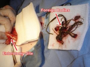
Gastrointestinal Foreign Bodies
A gastrointestinal foreign body is any non dietary material found within the digestive tract. These objects range from stones, coins, and string to toys, plant material and clothing. Pets of any age may swallow a foreign object, but most are consumed by puppies and kittens. Signs depend on the location of the foreign body, the amount of irritation and the degree of obstruction present. Common signs include anorexia, vomiting, abdominal discomfort, lethargy and the absence of stool. Diagnosis is afforded through accurate history, physical exam, abdominal imaging and endoscopy. Treatment usually involves endoscopic or surgical removal of the foreign body.
Gingival Hyperplasia
Gingival hyperplasia is an increase in the amount of gum tissue in the mouth. This condition is secondary to inflammation of the gum caused by dental tartar and/or infection. It presents as either a small isolated thickened nodule (epulis) or a generalized reddened gum. Diagnosis is via oral exam. Treatment includes dental cleaning, pain control, antibiotics and removal of excessive tissue.
Gingivitis
Gingivitis is an inflammation of the gums. The condition is caused by dental tartar, bacteria, foreign material (hair, plant material, food, etc.), irritants, etc. Gingivitis is a progressive disease. Slight reddening can progress to abscessation, ulceration, gingival hyperplasia, gingival recession, boney recession and tooth loss. Oral examination and dental radiographs are needed to diagnose and access the disease. Treatment consists of thorough dental cleaning, antibiotics and periodontal therapy.
Hemorrhagic Gastroenteritis
Hemorrhagic gastroenteritis is an inflammatory disease of the stomach and small intestine, characterized by acute vomiting and bloody diarrhea. Causes include infectious agents, immunological disorders, parasites, toxins, metabolic disorders and neoplasia. Diagnosis is afforded by complete blood counts, blood chemistries, laboratory tests, fecal exams, imaging studies and endoscopy with biopsy. Treatment usually consists of hospitalization, intravenous fluids, intravenous medication and dietary supplementation.
Histiocytic Ulcerative Colitis
Histiocytic Ulcerative colitis is a chronic inflammatory bowel disease of the large intestine of young (under 2 years of age) boxer dogs. The cause of the disease is unknown. Signs include severe, unresponsive, bloody, mucoid diarrhea and severe weight loss. Vomiting occurs in some dogs. Diagnosis is based on history, breed predisposition, and the endoscopic biopsy (positive periodic acid-Schiff (PAS) stain). Treatment is usually life long and includes anti-inflammatories, antimicrobials and a highly digestible diet.
Intestinal Obstruction
Intestinal obstruction is the blockage (partial or complete) of the normal passage of food through the intestinal tract. Causes include foreign bodies, anatomical anomalies, intestinal telescoping upon itself (intussusception), tumors, impacted food material, parasites and paralysis of the gut. Signs include vomiting, anorexia and abdominal pain. Diagnosis is afforded by history, physical exam, fecal exams, laboratory tests and abdominal imaging. Treatment ranges from medication, endoscopic extraction and/or diet to surgery.
Liver Disease
The liver performs many essential functions such as detoxifying poisons, processing drugs, metabolizing fats, proteins, carbohydrates and synthesizing proteins used throughout the body for growth, blood formation, blood clotting, metabolism, etc. Liver disease is any process by which the liver can no longer fully carry out its many functions and/or any process damaging the liver parenchyma. Causes of liver disease include degenerative processes, dysfunction secondary to anatomical disorders (eg. liver shunts), metabolic disorders, neoplasia, infectious diseases, inflammatory diseases, immunological disorders, trauma and toxins. Signs include anorexia, jaundice (yellow mucous membranes), vomiting, diarrhea, weight loss, poor coat, bleeding disorders, nervous disorders, shock and death. Diagnosis is afforded via complete blood counts, blood chemistries, laboratory tests, clotting panels, toxicological screens, serum bile acids, sulfated and nonsulfated urinary bile acids, ultrasonography, history, special imaging studies, histopathology (biopsy) and special immunological testing. Treatment may include special diet, immunomodulators(eg. ursodeoxycholic acid, prednisolone, mycophenolate, azathioprine, etc.), inhibitors of ammonia absorption, nutrients (eg. vitamine E, zinc acetate, s-adenosylmethionine), hepatoprotectants (eg. silibinin), omega 3 fatty acids and/or surgery.
Magaesophagus
Megaesophagus is a condition whereby the esophagus is dilated and cannot properly move food from the throat down to the stomach. Inhalation pneumonia is a common sequella due to pieces of ingesta remaining in the esophagus being inhaled into the lungs. The condition may be inherited (eg. Wire-haired Terriers and Miniature Schnauzers). Other causes include degenerative muscle diseases, neoplasia, immune disorders (eg. acetyl choline antibodies), hypothyroidism, trauma and toxins. Treatment aimed at the etiology may include immune therapy, thyroid hormone replacement, muscle receptor activators, surgery, elevating the pet’s food and water to aid flow to the stomach and/or treatment of any secondary problems (eg. inhalation pneumonia).
Malabsorption Syndrome
Malabsorption syndrome is a condition whereby the pet can not absorb digested nutrients from the small intestine into the bloodstream. Causes include immune disorders, inflammatory bowel disease, enzyme transport dysfunction, anatomical disorders, neoplasia, and various unknown causes (idiopathic). Possible signs are loose stools with a fetid order, anorexia, weight loss, bleeding disorders, weakness and poor hair coat. Diagnosis is afforded through clinical signs, physical exam, complete blood count, blood chemistries, absorption tests (eg. five sugar absorption, tagged nutrients, etc.), imaging studies, endoscopy and biopsy. Treatment is directed toward both the etiology of the problem as well as replenishment of lost nutrients.
Pancreatitis
The pancreas is an abdominal organ that lies next to the first part of the small intestine (duodenum) and stomach. It produces enzymes which break down food for digestion as well as insulin which regulates blood sugar.
Pancreatitis is an inflammation of the pancreas. Causes include ingestion of large amounts of fat, “rich” diets, infection, injury, or blockage of the pancreatic duct. Although it may occur in any individual, those most frequently stricken are overweight, middle-aged, female dogs.
Signs include pain, vomiting, depression, anorexia and occasionally shock and death. Complete blood counts, blood chemistries (including lipase and cPLI[canine pancreatic lipase immunoreactivity] or fPLI[feline pancreatic lipase immunoreactivity]), laboratory tests, abdominal imaging and physical exam are used to diagnose the condition. Treatment should be aggressive and long enough for complete recovery. This normally includes intravenous fluids, antibiotics, and no alimentation for an extended period; followed by a bland diet. Consequences of sustained pancreatitis may include actual autodigestion (enzymes destroying both the pancreas itself and surrounding organs!) and/or destruction of the insulin producing cells of the pancreas resulting in diabetes mellitus. Pets suffering from this condition should seek veterinary care immediately!
Periodontal Disease
Periodontal disease is a dynamic process that breaks down the structures surround teeth and hold them in their normal position. The disease begins with the accumulation of plaque (a clear combination of food particles, saliva and bacteria) upon the tooth surface down in the gingival sulcus (a canal between the tooth and gum). As plaque continues to accumulate, irritation of the gingiva (gums) produces inflammation (gingivitis). Gingivitis leads to bleeding of the gums. This bleeding mixes blood components with plaque producing tartar (calculus). As the process continues, the tartar works its way down the tooth root as well as out onto the visible surface of the tooth. Tartar and its contaminants can loosen teeth and spread infection leading to local abscess, as well as heart, liver, and kidney disease. Contaminants in contact with the lining of the mouth, can lead to tumor formation. Diagnosis of dental disease involves complete oral examination and dental radiographs. Treatment includes cleaning, polishing, root planing, gingival treatment and possible endodontic procedures.
Pharyngitis
Pharyngitis is an inflammation of the back of the throat (pharynx). Causes include infectious agents (eg. bacteria, viruses, fungi, etc.), periodontal disease, neoplasia, trauma, toxins and irritants. Signs may be redness of the mouth, reluctance to swallow, enlarged lymph nodes, coughing and/or blood-tinged saliva (hemapytalism). Diagnosis is possible through physical exam, complete oral exam, complete blood count, cytology, accurate history and cultures and sensitivities. Treatment may include antimicrobials, anti-inflammatories, possible removal of foreign bodies, surgery, dental work and special diet.
Protein Losing Enteropathy
Protein-losing enteropathy is a disease whereby proteins are lost through the intestine. Causes include impaired intestinal lymphatic drainage (eg. lymphangiectasia) that results in drainage of protein rich lymph into the bowel, neoplasia, inflammatory bowel disease, immune disorders, infectious diseases (eg. fungus) and disruption of the intestinal lining. Signs are weight loss, diarrhea, abdominal fluid (ascites) and/or edema (swelling of the limbs). Diagnosis is afforded through physical exam, complete blood count, blood chemistries, laboratory tests, abdominal fluid analysis, fecal exam, endoscopy with biopsy, immunological testing and imaging studies. Treatment may include special diets, antimicrobials, immune therapy, protectorants and/or corticosteroids.
Protosystemic Shunt
Portosystemic shunting is an abnormality of the liver’s blood vessels (portal system) whereby some or most of the blood from the intestines by-pass (shunt) the liver. The blood then goes directly into the general (systemic) circulation without the liver processing it. The consequences of this shunt are that blood ammonia levels rise after meals, which impairs brain function and may lead to seizures, coma and death. In addition, growth and general body functions are impaired. Portosystemic shunts may be present at birth (congenital) or develop later in life (acquired) secondary to liver disease. Signs of the disease include episodic weakness, ataxia, head pressing, disorientation, circling, pacing, behavioral changes, blindness, seizures, coma or death, stunted growth, vomiting, hypersalivation (especially in cats) and urinary crystals (urolithiasis). Diagnosis is afforded by physical exam, complete blood count, blood chemistries, imaging studies (ultrasonagraphy, contrast radiography) and/or exploratory surgery. Treatment includes surgery and medical management of the secondary clinical abnormalities (eg. seizures, vomiting, etc.)
Pyloric Stenosis/Pylorospasm
The pylorus is the short muscular passage from the stomach to the small intestine. Under normal conditions, the pylorus remains closed as the stomach churns and digests food. It then periodically opens in conjunction with the stomach as contractions pass food into the small intestine. Pylorospasm is when the pylorus fails to relax and allow food to pass from the stomach. Pyloric stenosis is a narrowing of the pylorus, also impeding the passage of food from the stomach. Pyloric stenosis is either congenital (present at birth) or acquired due to chronic infection, chronic hypertrophic pyloric gastropathy, neoplasia (eg. adenocarcinoma), inflammatory disease, trauma and secondary scarring. Pylorospasm may be due to metabolic disorders, neoplasia, infections (eg. bacterial, viral, fungal), inflammatory disease, trauma or toxins. Both conditions result in vomiting. The vomiting may be projectile. Other signs include anorexia and dehydration. Diagnosis is afforded by physical exam, complete blood count, blood chemistries, imaging studies, endoscopy, exploratory surgery and biopsy (histopathology). Treatment may consist of drugs that ameliorate gastric outflow (eg. metoclopramide, propulsid), acid blockers, antimicrobials, protectorants and/or surgery.
Rectal Prolapse
Rectal prolapse is the condition whereby a portion of the colon (lower digestive tract) turns inside out and protrudes from the anus. The prolapse results from straining due to diarrhea, constipation, neoplasia, inflammatory bowel disease, prostatitis, intestinal parasites, foreign objects and/or delivery of puppies or kittens. Clinically the disorder presents as a pink or red protrusion from the anus. Diagnosis is via physical exam. Treatment may include replacement or surgical resection of the prolapse, followed by eliminating the cause of the straining.
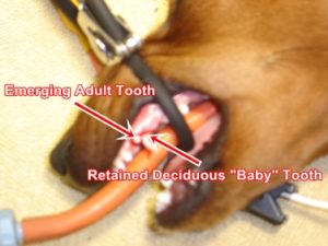
Retained Deciduous Teeth
Retained deciduous teeth are “baby teeth” that have not fallen out with the eruption of the corresponding permanent “adult” tooth. Causes include failed development of corresponding permanent teeth and/or misalignment of erupting and deciduous teeth. Either case prevents the dissolution of the deciduous tooth root and subsequent retainment of that tooth. Signs may include visualization of both corresponding teeth, malodor and/or oral pain. Diagnosis is afforded via physical exam and/or dental radiographs. Treatment is the extraction of the retained deciduous tooth as well as orthodontic intervention if warranted.
Salivary Cyst
Saliva from the salivary glands passes through ducts (passageways) to the mouth. Rupture of the duct allows saliva to accumulate into surrounding tissue. Causes include trauma, neoplasia and other obstructions to flow. Sign of the disorder is a soft, fluid filled cyst(sac) that has developed near the neck and/or jaw. Diagnosis is afforded by physical exam, aspiration, cytology, imaging studies and/or dye studies. The best treatment is surgical removal of the salivary gland forming the cyst. Recurrence is possible.
Spastic Colon
Spastic colon is an intermittent irritability of the colon (large bowel). Causes of the disorder include psychological stress, metabolic disorders, neoplasia, inflammatory bowel disease, bacterial overgrowth, infection (bacteria, virus, fungus, protozoa), parasites, foreign bodies, trauma and toxins. Signs include frequent passage of small amounts of soft stool that may include blood and/or mucous. Diagnosis is accomplished by fecal exam, physical exam (including digital exam), complete blood count, blood chemistries, laboratory tests (folate and cobalamin assays) and endoscopy with biopsy. Treatment is directed at the underlying disease. This may include antibiotics, endoscopic procedure (foreign body retrieval), surgery, high fiber diet, antimicrobials, sulfasalazine and/or stress management (physical and medical).
Stomatitis
Stomatitis is an inflammation of the mouth. Causes include metabolic disorders (eg. renal failure, diabetes mellitus, hypothyroidism), neoplasia, nutritional disorders (eg. niacin deficiency), infection (eg. bacteria, virus, fungal), periodontal disease, immune disorders (eg. systemic lupus erythamatosis, pemphigus complex, bulbous pemphigus), trauma and toxins (eg. acids, strong alkalis, petroleum distillates, drugs, etc.). Signs include red/sore mouth, bleeding gums, anorexia, foul odor, weight loss and poor condition. Diagnosis is afforded by a complete oral exam, biopsy, dental x-rays, complete blood counts, blood chemistries, laboratory tests, accurate history (eg. diet, exposure), culture and sensitivities. Treatment is directed at the underlying disease, which may include antimicrobials, dietary supplementation, complete dental cleaning and care, removal of foreign bodies, immunosuppressant therapy, cancer therapy and/or symptomatic therapy.
Tonsillitis
The tonsils are lymph tissue located on either side of the back of the throat. They function to destroy microorganisms entering the nose and throat. Tonsillitis is the inflammation of the tonsils. Causes of tonsillitis include chronic vomiting or regurgitation, chronic productive cough and chronic contamination of the nasopharynx (eg. severe periodontal disease). Signs of tonsillitis include cough, fever, retching, anorexia and lethargy. Diagnosis is afforded by oral exam (swollen with either red, hemorrhagic or white punctate, abscessed foci) and culture/sensitivity. Treatment is via antibiotic therapy and occasionally surgical removal of the tonsils.
Tooth Root Abscess
Tooth root abscesses commonly occur when a tooth fracture or wear allows exposure of the pulp cavity (root canal). This exposure allows bacterial access to the roots, surrounding bone other structures. The carnassial (“shearing”) tooth is most commonly affected in pets. (Fourth upper premolar in dogs; third upper premolar in cats.) Infections in this area may appear as a swelling or abscess below the eye. Other signs include pain, local swelling, reluctance to chew hard food and bleeding. A complete oral exam and dental radiographs (x-rays) are used to diagnose the condition. Treatment includes drainage of the infection, appropriate antibiotic treatment and either removal of the affected tooth or endodontic treatment.


